Amyloids
The conversion of proteins from a soluble into a fibrillar state is associated with a wide range of pathological conditions, including neurodegenerative diseases and systemic amyloidoses. The most prominent examples are Alzheimer's, Parkinson's and the prion diseases. However, also Diabetes type 2 is associated with the deposition of a hormone, hIAPP, in the pancreas of a diabetic patient. Amyloids are defined by the ability to bind to small molecules such as Thioflavin T or Congo Red. Their structures are characterized by a high degree of beta-sheet content. In the past years, it turned out that these aggregates have a tertiary structure and adopt a well defined "fold". However, it is still unclear how large the conformtional variations are and how this conformation affects cellular toxicity.
Structural Characterization of beta-amyloid peptides (Abeta)
Alzheimer's disease is the most common form of age-related neurodegenerative disorder. Abeta is obtained after processing of APP, the amyloid precursor protein. We study Abeta fibrils using solid-state NMR in order to better understand the mechanisms which lead to the formation of these aggregates.

Abeta fibril structure
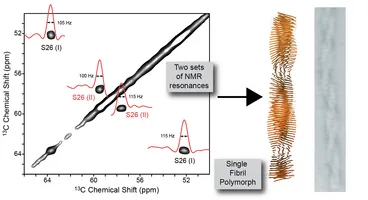
In the past few years, several structural models of Alzheimer's disease Abeta fibrils have been published (Petkova and Tycko, 2006; Paravastu and Tycko, 2008; Xiao and Ishii, 2015; Colvin and Griffin, 2016; Wälti and Riek, 2016). In these structural models, the basic fibril building block is composed of symmetric oligomers. In agreement with cryo-electron microscopy studies, we found that Abeta polymorphs can as well consist of asymmetric oligomers (Lopez del Amo et al., Angewandte Chemie 2012). We are currently working on the structural analysis of this particular amyloid polymorph. [image: Lopez del Amo et al., Angewandte Chemie 2012]
Abeta inhibtors
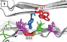
Non-steroidal anti-inflammatory drugs (NSAIDs) are known gamma-secretase modulators; they influence Abeta populations. NSAIDs are pleiotrophic and can interact with more than one pathomechanism. We demonstrated that the NSAID sulindac sulfide interacts specifically with Alzheimer disease Abeta fibrils. Sulindac sulfide does not induce drastic architectural changes in the fibrillar structure, but intercalates between the two beta-strands of the amyloid fibril and binds to hydrophobic cavities, which are found consistently in all analyzed structures (Prade et al., JBC 2015; Prade et al., Biochemistry 2016). Using chemical shift perturbation and 19F-13C distance restraints from REDOR experiments, we determine the binding site of these small molecules. The obtained results will allow to design more potent molecules for the treatment of the disease in the future. [image: Prade et al., JBC 2015]
Interactions between Small heat shock proteins and misfolding proteins
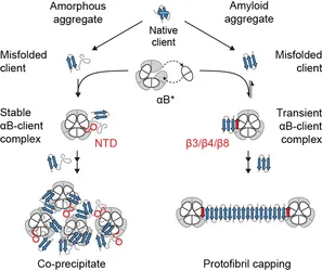
Small heat-shock proteins, including αB-crystallin (αB), play an important part in protein homeostasis, because their ATP-independent chaperone activity inhibits uncontrolled protein aggregation. Mechanistic details of human αB, particularly in its client-bound state, have been elusive, in part due to the high molecular weight and the heterogeneity of these complexes. We have shown using NMR spectroscopy, that the αB complex is assembled from asymmetric building blocks. Interaction studies demonstrated that the fibril-forming Alzheimer's disease Aβ1–40 peptide preferentially binds to a hydrophobic edge of the central β-sandwich of αB. In contrast, the amorphously aggregating client lysozyme is captured by the partially disordered N-terminal domain of αB. We suggest that αB uses its inherent structural plasticity to expose distinct binding interfaces and thus interact with a wide range of structurally variable clients (Mainz et al., Nature Struct. Mol. Biol. 2015). [image: Mainz et al., Nature Struct. Mol. Biol. 2015]
Soluble Protein Complexes
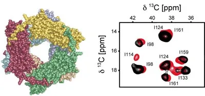
The relaxation rate of a nucleus in solution-state NMR is determined by the tumbling rate of the molecule. Typically, line widts increase for larger molecules, making it increasingly difficult to characterize molecules which are larger than 100 kDa. Solid-state NMR is not limited by tumbling correlation time, as the molecules are immobilized. Therefore, the experimental line width is independent of the size of the molecule. Immobilization of the molecule can be achieved by crystallization, but also by just increasing the viscosity of the solution. This approach was demonstrated using of the small heat shock protein αB-crystallin. This techniques opens new perspectives for the investigation of large protein complexes, as ligands do not have to be co-precipitated to study protein-protein interactions.
[image: MAS NMR 13C,13C correlations carried out for the small heat shock protein αB-Crystallin (600 kDa) in solution. Mainz et al., J. Am. Chem. Soc. 2009]
Proteasome complexes
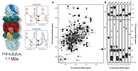
Various cellular processes are regulated by large molecular machines. The proteasome degradation machinery is essential for cellular protein homeostasis and viability. We could show that a combination of magic-angle spinning (MAS) and proton detection enables us to obtain sequential backbone assignments for a megadalton archaeal proteasome complex without the need for protein crystallization or precipitation. Rotational diffusion of the solute protein complex is impaired by MAS-induced reversible sedimentation. The approach permits exploration of the structure, dynamics and interactions of supramolecular assemblies in the megadalton regime (Mainz et al., Angewandte Chemie 2013)
Ribosomal Complexes
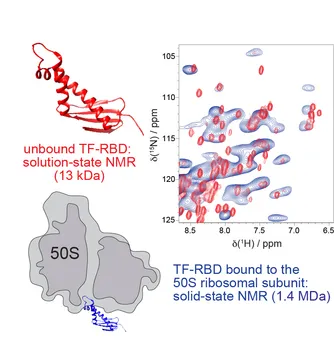
Co-translational protein folding is not yet well understood despite the availability of high-resolution ribosome crystal structures. We could demonstrate in preliminary experiments that the non-mobile regions of a prokaryotic ribosomal complex can be studied using solid-state NMR. This large asymmetric protein complex (1.4 MDa) becomes accessible by the combined use of proton detection and high MAS frequencies (60 kHz). These experiments open new perspectives for the understanding of the mechanism of large molecular machineries (Barbet-Massin, Angewandte Chemie 2015)
Solid-State NMR Methods
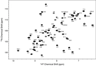
So far, solid-state NMR experiments that measure distances or torsion angles between heteroatoms, are restricted to doubly labeled peptide or protein samples. We develop experiments that are suitable to characterize the structure of uniformly labeled peptides proteins. Furthermore, we develop labeling concepts that allow to detect protons with high sensitivity. These strategies rely on perdeuteration of the protein with backsubstitution of the exchangeable deuterons with protons from the solvent. The spectrum above shows an HSQC spectrum of a-spectrin SH3 domain obtained in the solid-state. The obtainable resolution is comparable to a the resolution that is achieved in solution-state NMR for an intermediate sized protein. (Chevelkov et al., Angewandte Chemie Int. Edt. 2006; Linser et al., J. Am. Chem. Soc. 2010)
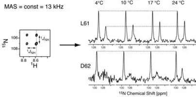
One focus of the research is the quantification of dynamics. Solid-state NMR is like no other method suited to describe dynamics. In solution, most of the relaxation is due to overall tumbling. By contrast, relaxation in the solid-state is only due to local structural fluctuations. The 1D-15N spectra above are cross sections along the 15N dimension in a HSQC experiment that was recorded without scalar decoupling in t1. Accordingly, signals are split into doublets. Intensities of the 15N-1H(α) and 15N-1H(β) spin states are assymetric due to dipole-CSA cross correlated relaxation. Differential relaxation of the multiplet components is due to nano-sec local structural fluctuation.
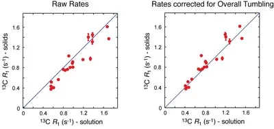
In collaboration with Prof. Nikolai Skrynnikov, Purdue University, we compare the dynamic properties of the α-spectrin SH3 domain in the crystalline state and in solution. We find that the motional properties are highly similar. This opens new perspectives for the dynamic characterization of a protein. The image shows the correlation of methyl 13C-R1 relaxation rates for the α-spectrin SH3 domain in solution and in the solid-state. Even without the correction term which takes into account overall tumbling, the relaxation rates are very similar (Agarwal et al., J. Am. Chem. Soc. 2008).
